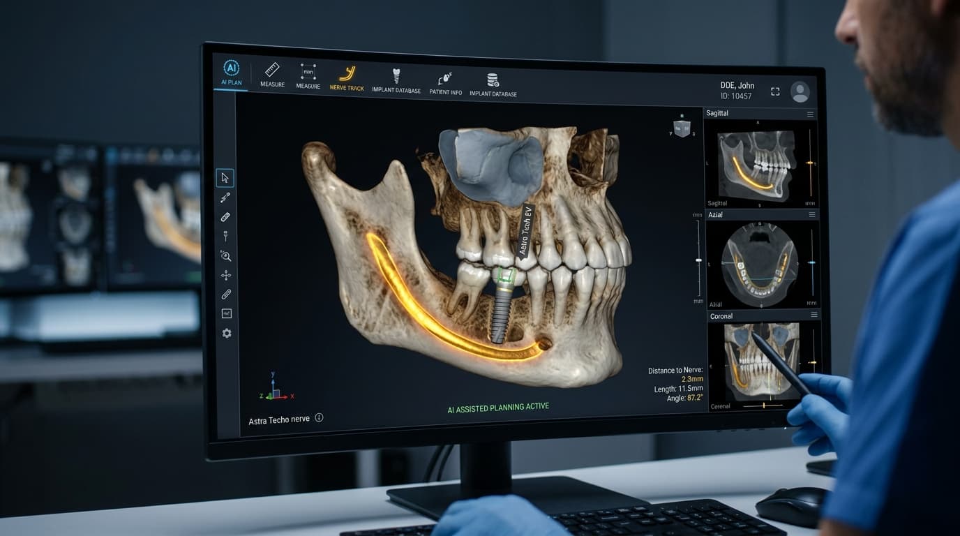
More implant patients now see their treatment plan before they ever sit in the chair. The surgeon pulls up a 3D model of your jaw, the nerve running through it is highlighted, the sinus floor is shaded, and a virtual implant sits in the bone at a precise angle. Increasingly, the first draft of that picture isn't drawn by a person. AI software produces it from your CBCT scan in a matter of seconds.
What patients rarely hear is how that software learned to find a nerve in the first place. Every AI model that reads a dental CBCT scan was trained on thousands of scans that people outlined by hand, structure by structure, slice by slice. I've spent years around that kind of work, and the honest answer is that the AI is only as careful as the people who labeled its training data. That's why serious developers rely on specialized medical data annotation teams with radiologists checking the work, rather than treating dental scans like generic image tagging.
So if you're considering dental implants and your clinic mentions AI-guided planning, here's what's actually happening behind the screen: what the technology does well, where it can slip, and what to ask before you book.
What Is AI-Guided Dental Implant Planning?
Implant planning has always meant answering the same handful of questions. How much bone is there? Where are the nerve and the sinus? What implant position will best support the final crown? AI-guided planning answers the first two much faster. The software takes your cone beam CT (CBCT) scan, automatically segments the anatomy (teeth, jawbone, nerve canal, sinus) and gives your surgeon a ready-to-review 3D model. Some systems also suggest an implant size and position based on the available bone and the planned crown.
From there, the plan can be turned into a 3D-printed surgical guide: a thin template that fits over your teeth or gums and steers the drill to the planned spot, depth, and angle. That's what's usually meant by guided implant surgery. The AI speeds up the planning, the guide carries the plan into your mouth, and the surgeon stays responsible for both.
One thing worth clearing up early: "AI-guided" doesn't mean a robot places your implant. In nearly every practice, AI is a drafting tool. Your surgeon reviews, corrects, and signs off on every measurement.
Why Does Dental Implant Planning Start With a CBCT Scan?
A regular panoramic X-ray is flat. It shows the height of your jawbone reasonably well, but it tells your dentist very little about the bone's width, and width is often what decides whether an implant fits. A CBCT scan captures your jaw in three dimensions, so the surgeon can look at the bone in cross-section at the exact spot where the implant will go.
That difference shows up in real outcomes. A 2024 study in Dentistry Journal that reviewed 200 implant cases found that every patient who had a CBCT scan during planning received their implant as scheduled. In about 7% of the cases planned without one, surgery was stopped partway through, mostly because there wasn't enough bone or the implant couldn't be stabilized.
CBCT is also the raw material for AI. The scan is made of hundreds of thin image slices stacked into a volume of tiny 3D pixels called voxels. Every voxel is a data point, and the 3D model is only trustworthy if those data points are interpreted correctly.
What Does AI Actually Look For on Your CBCT Scan?
When planning software reads your scan, it's doing what a radiologist does, just much faster. It's hunting for the structures that make or break an implant:
- The inferior alveolar nerve canal. This nerve runs through the lower jaw and supplies feeling to your lower lip and chin. It's the structure implant surgeons respect most.
- The mental foramen. The small opening where that nerve exits the jaw near the premolars.
- The maxillary sinus. The air space above your upper back teeth, which often limits how much bone is available there.
- Bone height, width, and density. Including the thin outer layers of cortical bone that give an implant its initial stability.
- Neighboring tooth roots. An implant placed at the wrong angle can contact the root next to it.
Identify those correctly, and the plan has the safety margins it needs. Miss them by a couple of millimeters, and a plan can look perfect on screen while pointing toward a nerve.
What Is CBCT Data Annotation, and Why Should Patients Care?
Before any AI can recognize a nerve canal, it has to be shown what one looks like, thousands of times, on thousands of different jaws. That teaching material comes from data annotation: trained specialists open real, anonymized CBCT scans and mark every relevant structure, voxel by voxel.
In practice, it looks like this. An annotator traces the nerve canal on one slice, then the next, then the next, following it as it curves through the jaw and sometimes fades into the surrounding bone. They outline the sinus floor, the bone borders, each tooth root. Then a radiologist reviews the work. On well-run projects, when two experts disagree about where a boundary sits, a senior reviewer makes the final call. That's called adjudication, and it's exactly the step that cheaper pipelines tend to skip.
Why should this matter to you as a patient? Because a model learns its labels, mistakes included. If the training data consistently placed the nerve canal half a millimeter too high, the AI will learn to place it half a millimeter too high, on your scan as well. Nobody can spot that kind of built-in error later by looking at the software's confident 3D render.
Oleksandr Chyrkov, a board-certified radiologist and Quality Manager at medDARE with more than 14 years in CT and MRI diagnostics, puts it this way:
"On a CBCT scan, the mandibular canal can disappear for several slices in patients with low bone density, and that is exactly where a model learns to guess. When we review annotations, we don't only ask whether a contour looks right. We ask whether a second radiologist, scrolling through the same volume independently, would draw the same nerve. If two experts disagree by a millimeter, that millimeter goes to adjudication, because in implant planning a millimeter is the whole safety margin." — Oleksandr Chyrkov, Board-Certified Radiologist and Quality Manager, medDARE
There's a privacy side too. Training scans come from real patients, so responsible datasets are anonymized before annotation, with names, dates, and other identifying details stripped from the image files, and they're handled under frameworks such as HIPAA in the United States and GDPR in Europe.
How Accurate Is AI in Dental Implant Planning?
In research settings, the reported benchmark results are genuinely impressive. One of the largest studies to date, published in Nature Communications, trained an AI system on 4,938 CBCT scans from 4,215 patients across 15 centers. It segmented teeth and alveolar bone about as accurately as experienced radiologists and did it roughly 500 times faster, including on difficult cases with dental abnormalities.
Notice what made that possible: a large, varied dataset from many clinics and scanners, carefully labeled. The accuracy figures in AI papers describe how closely a model matches its reference labels. When those labels are excellent and diverse, a high score means something. When they're inconsistent, or all come from one type of patient, a high score can hide real problems.
For the surgery itself, guided placement is generally more precise than freehand placement. Systematic reviews typically report average deviations of roughly one to one and a half millimeters between the planned and final implant position. That's precisely why careful surgeons still build a safety buffer, commonly around 2 mm, between the implant and the nerve canal.
Where Can AI Implant Planning Go Wrong?
From what I've seen, AI rarely struggles with textbook cases. It struggles with jaws that don't look like its training data. The usual trouble spots are:
- Metal artifacts. Crowns, large fillings, and existing implants throw bright streaks across the scan that can hide or distort nearby anatomy.
- Low-density bone. In softer bone, the nerve canal may lack a clear border, so its path has to be inferred rather than seen.
- Anatomical variations. Some people have a nerve that loops forward before it exits the jaw, a canal that splits in two, or extra small openings. These are common enough to matter and rare enough to be underrepresented in training data.
- Previous surgery. Bone grafts and healing extraction sockets can look very different from untouched bone.
- Scan quality. Patient movement or low-resolution settings blur the fine details the software depends on.
Each of these has a data-side fix: the training set needs enough of these hard cases, labeled by people who know what they're looking at. Each has a clinical fix as well: your surgeon should check the AI's segmentation slice by slice in the critical areas, not just glance at the 3D model.
Is AI Implant Planning Software Regulated?
In the United States, software that helps diagnose conditions or plan treatment is generally regulated as a medical device. The FDA maintains a public list of AI-enabled medical devices authorized for marketing, which had grown past 1,600 devices as of September 2026, with radiology accounting for the largest share. Dental imaging software appears there too. In Europe, comparable software falls under the Medical Device Regulation and needs a CE mark.
Clearance tells you the software met a standard for its intended use. It doesn't mean every plan it produces is correct, which is why your clinician's review remains part of the process.
Does AI Replace Your Implant Surgeon?
No, and good clinics don't pretend otherwise. AI takes over the tedious part: tracing anatomy across hundreds of slices. The judgment calls stay human. Should you have a bone graft first? Would a shorter implant be safer than a sinus lift? How should the final crown sit in your bite? How do your medical history and healing habits change the plan? A treatment plan is only as good as the person who signs it.
What Should You Ask Before AI-Guided Implant Surgery?
If you're comparing clinics, especially abroad, these questions separate a genuinely modern workflow from a marketing line:
- Will I have a CBCT scan, and will you walk me through it?
- Is AI used in my planning, and who reviews its output?
- Will you use a surgical guide, and is it fully or partially guided?
- How much distance does my plan leave from the nerve or sinus?
- Can I get a copy of my CBCT scan in DICOM format?
A confident implant surgeon will be happy to answer all of these. If "it's AI-guided" is the entire answer, keep asking.